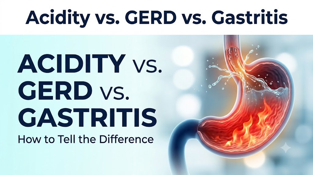
Almost everyone has experienced that familiar, uncomfortable sensation: a fiery heat creeping up the chest after an indulgent late-night dinner, a sour taste filling the back of the mouth, or a gnawing, empty ache right below the ribs. In casual conversation, these sensations are almost always grouped under a single, catch-all umbrella: “acidity.”
In households and workplaces across urban centers, the standard response to this discomfort is predictable. People pop an over-the-counter antacid, drink a glass of cold milk, dissolve effervescent salts in water, or sip on traditional herbal remedies. When the burning subsides an hour later, the problem is considered solved.
“Burning in Stomach” ──► Assumed to be “Simple Acidity” ──► Daily Antacids
THE REALITY:
• Acidity = An occasional physiological symptom of acid irritation.
• GERD = A mechanical failure of the valve between the stomach and the esophagus.
• Gastritis = Structural inflammation, erosion, or infection of the stomach wall.
Relying on antacids to continuously suppress upper abdominal distress is like silencing a smoke detector while a fire smolders behind the wall. While occasional acidity is an episodic physiological reaction, Gastroesophageal Reflux Disease (GERD) and Gastritis are distinct, progressive medical conditions with unique anatomical targets, completely different physiological mechanisms, and potentially severe long-term complications if left untreated. Understanding the differences between these three conditions is not merely a matter of academic trivia—it is essential for protecting the lining of your esophagus and stomach from irreversible damage.
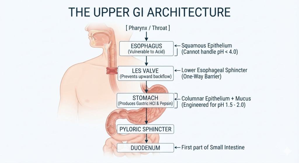
To understand why these conditions feel similar yet behave differently, we must examine the architectural layout of the upper gastrointestinal (GI) tract.
The Stomach: An Acid-Resistant Fortress
The human stomach is a muscular organ specifically engineered to function in an intensely acidic bath. Its parietal cells produce hydrochloric acid (HCl) alongside the digestive enzyme pepsin. Gastric juice has a pH ranging between 1.5 and 2.0—an acidity level potent enough to dissolve plant fibers, break down tough meat proteins, and destroy ingested pathogens.
The stomach survives this caustic fluid through a multi-layered defense mechanism known as the Gastric Mucosal Barrier:
- The Bicarbonate-Rich Mucus Layer: Specialized foveolar cells secrete a thick gel of water, glycoproteins, and bicarbonate ions. This creates a protective, neutral pH gradient directly over the stomach tissue.
- Tight Epithelial Junctions: Surface cells are packed closely together, preventing acid ions from leaking into deeper layers of tissue.
- Microvascular Blood Flow: A network of capillaries rapidly delivers oxygen and micronutrients while clearing any acid that penetrates the surface.
- Prostaglandin Signaling: Tissue messengers (especially Prostaglandin) continuously stimulate mucus production and repair minor daily wear and tear.
The Esophagus: An Unprotected Corridor
Above the stomach sits the esophagus—a 10-inch muscular tube tasked with propelling food downward via coordinated peristaltic contractions. Unlike the stomach, the esophagus is lined with delicate stratified squamous epithelium. It possesses no thick mucus coat and has zero natural defenses against strong acids or digestive enzymes.
The Lower Esophageal Sphincter (LES): The Gatekeeper
Under normal circumstances, the corrosive contents of the stomach are prevented from splashing into the vulnerable esophagus by the Lower Esophageal Sphincter (LES). This ring of smooth muscle, located where the food pipe joins the stomach, acts as a high-pressure, one-way physiological valve.
It relaxes briefly for 5 to 8 seconds during swallowing to let food enter the stomach, then immediately clamps shut, maintaining a high-pressure seal against the churning acidic liquid below.
2. Acidity: The Occasional, Acute Disturbance
What Is It?
“Acidity” is not a recognized standalone medical diagnosis in modern gastroenterology. Rather, it is a colloquial umbrella term used to describe transient, self-limiting heartburn, sour belching, or mild stomach discomfort triggered by temporary lifestyle, dietary, or emotional factors.
Pathophysiology
In acute acidity, the underlying mucosal anatomy of the stomach and esophagus remains structurally sound. There are no persistent ulcers, no tissue erosion, and no permanent mechanical defect in the LES valve.
Instead, the digestive system is simply overwhelmed:
- Volume Overload: An unusually heavy or high-fat meal takes longer to digest, causing food to sit in the stomach for several hours.
- Pressure Buildup: The expanded stomach increases intragastric pressure, pushing up against the LES.
- Transient Valve Slippage: The valve experiences a brief, normal physiological relaxation (known as a Transient Lower Esophageal Sphincter Relaxation, or TLESR). A small splash of acid escapes into the lower esophagus, triggering sensory nerve endings.
Typical Causes and Triggers
- Large, rich, deep-fried, or heavily spiced meals.
- Lying flat or slouching on a couch immediately after eating.
- Drinking carbonated sodas, excessive coffee, or alcohol (which directly relaxes the LES muscle).
- Severe, short-term psychological stress triggering temporary stomach muscle tension.
- Infrequent, erratic meal times resulting in concentrated acid secretions churning on an empty stomach.
Symptom Profile and Behavior
- Frequency: Occasional (once or twice a month, typically linked to a specific dietary indulgence).
- Duration: Short-lived, resolving within 30 to 90 minutes.
- Response to Simple Remedies: Clears rapidly after drinking water, standing upright, or taking a single over-the-counter antacid tablet.
- Absence of Progressive Symptoms: Does not cause difficulty swallowing, persistent vomiting, unexplained weight loss, or chronic morning hoarseness.
3. GERD (Gastroesophageal Reflux Disease): The Mechanical Failure
What Is It?
When acid reflux stops being an occasional post-dinner inconvenience and transforms into a chronic, recurring event, it crosses the threshold into Gastroesophageal Reflux Disease (GERD).
GERD is a chronic, progressive condition where stomach contents persistently back up into the esophagus, causing troublesome symptoms or macroscopic mucosal tissue damage.
| Feature | Normal Valve Function | GERD: Mechanical Failure |
|---|---|---|
| Esophagus Status | Content stays clear; lining remains unexposed and healthy. | Acid surges upward, causing chemical irritation or tissue damage. |
| LES Valve State | Tight Valve; acts as a secure, high-pressure one-way barrier. | Incompetent Valve; transiently relaxes, weakens, or fails to close. |
| Stomach Status | Acid stays contained within the protective, mucus-lined chamber. | Acid escapes freely due to a lack of mechanical resistance. |
Pathophysiology: Why the Gate Fails
Unlike simple acidity, GERD is primarily a mechanical and neuromuscular failure, not merely an issue of having “too much acid.” The barrier holding down stomach contents fails due to one or more factors:
1. Chronic Hypotensive Lower Esophageal Sphincter (Weak LES)
The resting pressure of the LES is normally between 10 and 45 mmHg. In patients with GERD, this basal pressure often drops below 10 mmHg. The valve stays loose and partially open, allowing digestive juices to splash upward every time the stomach contracts.
2. Hiatus Hernia (Anatomical Distortion)
The diaphragm muscle normally wraps snugly around the lower esophagus, providing an external “pinch-cock” muscular squeeze that reinforces the internal LES valve. In a Hiatus Hernia, the opening in the diaphragm (the hiatus) widens, allowing the upper section of the stomach to slide upward into the chest cavity. This completely separates the internal LES from the diaphragm’s external support, severely impairing anti-reflux defense.
3. Delayed Gastric Emptying (Gastroparesis)
If the stomach empties food into the small intestine sluggishly, prolonged pressure stretches the stomach wall, triggering frequent, prolonged valve relaxations.
4. Impaired Esophageal Acid Clearance
Normally, when acid splashes into the esophagus, secondary peristaltic clearing waves push the fluid back down, while swallowed saliva (rich in bicarbonate) neutralizes any remaining acidity. In chronic GERD, esophageal clearing motility often weakens, allowing acid to linger against the delicate lining for extended periods.
The Atypical and Silent Faces of GERD
While classic heartburn (pyrosis) and sour regurgitation are common, GERD frequently presents with “extra-esophageal” or atypical symptoms that can confuse patients and delay diagnosis:
ATYPICAL MANIFESTATIONS OF GERD:
• Chronic Nighttime Cough & Throat Clearing: Acid micro-droplets aerosolize and irritate vocal cords (Laryngopharyngeal Reflux / LPR).
• Dental Enamel Erosion: Acid gases dissolve protective enamel, leading to unexplained cavities and extreme tooth sensitivity.
• Adult-Onset Asthma / Wheezing: Acid aspiration triggers bronchial spasms
• Globus Sensation: Persistent feeling of a “lump” in the throat.
• Non-Cardiac Chest Pain: Squeezing chest discomfort mimicking angina
4. Gastritis: The Mucosal Fire Within
What Is It?
While GERD involves acid splashing upward out of the stomach, Gastritis is a problem located entirely within the stomach itself.
Gastritis is defined histopathologically as the inflammation, erosion, or irritation of the protective gastric mucosal lining.
| Feature | Healthy Gastric Mucosa | Active Gastritis |
|---|---|---|
| Mucus Barrier | Thick, glistening mucus layer that continuously neutralizes acid. | Broken mucus barrier that exposes the delicate tissue underneath. |
| Tissue Appearance | Intact cellular lining with a uniform, pink, and healthy appearance. | Fiery red inflammation accompanied by microscopic hemorrhages. |
| Acid Protection | Protected from deep acid damage by robust cellular tight junctions. | Raw erosions & ulcers form as gastric juices eat into the stomach wall. |
In gastritis, the problem is not necessarily that the stomach produces excessive acid. Instead, the protective mucus barrier is compromised or worn away, allowing normal stomach acid to come into direct contact with vulnerable stomach tissue, causing raw inflammation.
Primary Drivers of Gastritis
1. HELICOBACTER PYLORI (H. Pylori) INFECTION
• Spiral bacterium that burrows into the stomach’s protective mucus layer.
• Produces urease, creating ammonia that damages surrounding cells.
• Drives chronic, long-term inflammation across the gastric wall.
2. NSAID & PAINKILLER OVERUSE
• Medications like Ibuprofen, Diclofenac, Naproxen, and Aspirin.
• Systemically block COX-1 enzymes, stopping protective prostaglandin flow.
• Leaves the stomach lining vulnerable to acid digestion within days.
Other significant clinical causes include:
- Alcohol Abuse: Ethanol directly dissolves the gastric mucus layer, causing acute erosive or hemorrhagic gastritis.
- Autoimmune Gastritis: The body’s immune system produces autoantibodies that attack acid-producing parietal cells, leading to Vitamin B12 deficiency (pernicious anemia).
- Severe Physiological Stress: Critical illness, extensive burns, or major surgery can reduce protective blood flow to the stomach lining, triggering acute stress erosions.
- Bile Reflux: The pyloric valve at the bottom of the stomach malfunctions, allowing caustic, alkaline bile from the small intestine to wash backward into the stomach.
Acute vs. Chronic Gastritis
- Acute Gastritis: Develops rapidly, causing sudden, sharp, severe upper abdominal pain, nausea, and vomiting. It is typically triggered by heavy alcohol intake, food poisoning, or a burst of high-dose painkillers. Once the trigger is removed, the mucosal lining can heal relatively quickly.
- Chronic Gastritis: Progresses quietly over months or years, often driven by untreated H. pylori infection. It causes low-grade, nagging discomfort, early fullness, or mild nausea. Over time, chronic inflammation can cause the stomach lining to thin and lose its gland structure (Atrophic Gastritis), significantly increasing the risk of peptic ulcers and gastric tumors.
5. Comprehensive Comparison: Acidity vs. GERD vs. Gastritis
| Clinical Feature | Acidity | GERD | Gastritis |
|---|---|---|---|
| Primary Problem | Occasional acid surge due to diet/lifestyle choices. | Weak or failing lower esophageal sphincter (LES) valve. | Broken or inflamed protective stomach wall lining. |
| Pain Location | Behind the breastbone or high upper abdomen. | Lower chest, behind the sternum; moves upward. | Upper central abdomen (epigastrium), just below the ribs. |
| Pain Character | Mild, transient, warm burning sensation. | Sharp, fiery burning shooting upward into the throat. | Gnawing, aching, raw, hollow hunger-like pain. |
| Food Relationship | Appears 1–2 hours after eating a heavy meal. | Worsens when lying down or bending over after meals. | Pain often changes immediately after food intake. |
| Characteristic Signs | Sour burp, fleeting warm chest sensation. | Sour regurgitation, chronic dry cough, lump in throat, hoarseness. | Early fullness, nausea, loss of appetite, persistent bloating. |
| Underlying Mucosa | Completely normal, undamaged lining. | Esophageal erosions, ulcers, or Barrett’s esophagus. | Gastric mucosal edema, erythema, or stomach erosions. |
| Standard Care | Lifestyle adjustments and occasional antacids. | Long-term acid control, prokinetics, or surgical intervention. | Eradicate H. pylori, stop NSAIDs, protect the stomach wall. |
6. The Dangers of Chronic Self-Medication
In many households, over-the-counter antacids and Proton Pump Inhibitors (PPIs like omeprazole, pantoprazole, and rabeprazole) are treated like candy. Whenever stomach distress strikes, a pill is taken.
While these medications are valuable clinical tools when prescribed properly, relying on them for unmonitored self-treatment carries serious medical risks.
1. Masking Early Cancers and Precancerous Lesions
Stomach and esophageal cancers often present in their earliest, most curable stages with simple symptoms: mild indigestion, early fullness, or vague chest burning. Masking these symptoms with acid suppressants without an endoscopic evaluation can delay diagnosis until the disease reaches an advanced stage.
2. Hypochlorhydria and Poor Nutrient Absorption
Stomach acid is not an evolutionary accident—it is essential for breaking down and absorbing vital nutrients. Artificially suppressing stomach acid for months or years impairs the absorption of:
- Vitamin B12: Requires acid and intrinsic factor for separation from food proteins (deficiency causes peripheral neuropathy and anemia).
- Non-Heme Iron: Needs an acidic pH to convert into an absorbable form ($Fe^{2+}$); chronic suppression leads to refractory iron deficiency.
- Calcium and Magnesium: Reduced absorption can contribute to bone density loss (osteopenia and osteoporosis) and muscle cramping.
3. Small Intestinal Bacterial Overgrowth (SIBO) and Gut Infections
Gastric acid acts as a primary sterilization barrier against trillions of microbes ingested daily in food and water. When stomach acid is neutralized long-term, swallowed bacteria survive and colonize the small intestine, leading to SIBO (Small Intestinal Bacterial Overgrowth), chronic bloating, and increased risk of Clostridioides difficile intestinal infections.
7. What Happens if They Are Left Untreated?
Ignoring persistent upper GI symptoms allows localized inflammation to develop into structural complications.
Complications of Untreated GERD
Chronic Reflux ──► Erosive Esophagitis ──► Peptic Stricture ──► Barrett's Esophagus ──► Adenocarcinoma
- Erosive Esophagitis: Deep ulcers form in the esophageal lining, leading to spontaneous bleeding and severe, painful swallowing.
- Peptic Strictures: Ongoing acid burns trigger fibrous scar tissue formation. Over time, the lower esophagus narrows, making it difficult or impossible for solid food to pass into the stomach.
- Barrett’s Esophagus: In response to chronic acid washing over the lower esophagus, the body attempts to protect itself by replacing delicate squamous cells with specialized, acid-resistant intestinal columnar cells (metaplasia). While this reduces burning sensations, Barrett’s tissue is precancerous, carrying a significantly elevated risk of transforming into Esophageal Adenocarcinoma.
Complications of Untreated Gastritis
- Peptic Ulcer Disease: Mucosal erosions eat deep into the muscular wall of the stomach, exposing underlying blood vessels.
- Upper GI Hemorrhage: An eroded vessel can burst, resulting in vomiting fresh red blood (hematemesis) or passing jet-black, sticky stools (melena).
- Gastric Perforation: A deep ulcer burns entirely through the stomach wall, leaking acidic gastric contents into the abdominal cavity—a life-threatening surgical emergency.
- Gastric MALT Lymphoma and Carcinoma: Long-standing chronic atrophic gastritis driven by H. pylori is an established risk factor for gastric cancer.
8. When to See a Gastroenterologist: The Alarm Red-Flag Checklist
While occasional indigestion can be monitored with dietary changes, certain alarm symptoms require immediate clinical evaluation. If you experience any of the signs below, stop self-medicating and consult a gastroenterologist promptly:
- Dysphagia: Food catches, sticks, or fails to go down normally.
- Odynophagia: Sharp, burning pain experienced specifically while swallowing food or liquids.
- Unintentional Weight Loss: Dropping pounds significantly without active dieting or lifestyle changes.
- Persistent Vomiting: Continuing inability to keep food or liquids down over a period of time.
- Hematemesis: Vomiting bright red blood or dark “coffee-ground” material indicating upper GI bleeding.
- Melena: Passing foul-smelling, jet-black, tar-like stools caused by digested blood.
- Unexplained Anemia: Low hemoglobin levels accompanied by chronic fatigue, pale skin, or weakness.
- Palpable Abdominal Mass: Finding a new lump, hardness, or localized swelling in the stomach area.
- Late-Onset Symptoms: Developing severe heartburn or indigestion for the first time over the age of 45–50.
9. Modern Diagnostic Pathways in Gastroenterology
A gastroenterologist relies on precise diagnostic tools to evaluate the upper digestive tract rather than guessing based on symptoms alone:
1. High-Definition Upper GI Endoscopy (Gastroscopy)
- How it works: A thin, flexible tube with a high-definition digital camera and LED light is gently guided down the esophagus, into the stomach, and through to the duodenum.
- Patient experience: Performed under short-acting, conscious sedation. Patients rest comfortably for 8 to 10 minutes and wake up without pain, choking, or memory of discomfort.
- What it sees: Provides direct visualization of esophageal erosions, hiatus hernias, Barrett’s tissue, active gastritis, and peptic ulcers.
- Biopsy capabilities: The specialist can pass micro-forceps through the scope to take small, painless tissue samples, confirming H. pylori infection or ruling out precancerous changes.
2. Testing for Helicobacter Pylori
- Rapid Urease Test (RUT): A small biopsy taken during endoscopy is placed in a urea broth; an immediate color change confirms active urease-producing bacteria.
- Urea Breath Test (UBT): The patient swallows a specialized, harmless carbon-labeled urea solution. If H. pylori is present, it breaks down the urea, releasing labeled carbon dioxide that is detected in exhaled breath.
3. 24-Hour Esophageal pH-Impedance Testing
Considered the gold standard for diagnosing elusive GERD cases. A tiny, hair-thin catheter is placed in the lower esophagus for 24 hours to measure the exact frequency, timing, and height of acid and non-acid reflux episodes, matching them against the patient’s daily symptoms.
4. High-Resolution Esophageal Manometry
Uses pressure sensors to assess the muscular strength and coordination of the esophagus, measuring the exact resting squeeze pressure of the LES to evaluate valve competence.
Medical Interventions Under Specialist Care
- For Acute/Chronic Gastritis:
- If H. pylori is detected, a strict 14-day course of Bismuth-Based Quadruple Therapy (two targeted antibiotics, a bismuth mucosal protector, and a high-potency acid suppressant) is prescribed to eradicate the infection completely.
- If painkiller-induced, offending medications are stopped and replaced with stomach-sparing alternatives alongside sucralfate mucosal suspensions to allow the lining to heal.
- For Chronic GERD:
- Structured cycles of acid suppressants allow inflamed esophageal tissue to regenerate.
- Prokinetic agents may be used to improve stomach emptying and tighten the lower esophageal valve.
- For patients with severe hiatus hernias or valves that fail to close, minimally invasive procedures (such as Laparoscopic Nissen Fundoplication or Transoral Incisionless Fundoplication [TIF]) can reconstruct a physical barrier against reflux.
Foundational Lifestyle and Dietary Modifications
- Follow the 3-Hour Rule: Never lie flat within three hours of your last meal. Give your stomach time to empty into the small intestine while gravity keeps digestive juices where they belong.
- Elevate the Head of Your Bed: Raising the head of your bed frame by 6 inches prevents nighttime acid backflow. (Note: Stacking extra pillows under your head bends your neck without elevating your torso, which compresses the abdomen and worsens reflux.)
- Adjust Meal Sizes: Eating large meals expands the stomach and forces the LES open. Switching to four smaller, well-spaced meals throughout the day reduces intragastric pressure.
- Identify Personal Triggers: While common triggers include fried foods, chocolate, caffeine, citrus, peppermint, and alcohol, keeping a food diary helps pinpoint the specific ingredients that affect you.
- Manage Abdominal Pressure: Excess visceral fat presses directly against the stomach, pushing contents upward against the valve. Gradual, sustained weight reduction often resolves mild to moderate GERD without long-term medication.
Navigating upper digestive discomfort comes down to recognizing the clear differences between these three conditions:
- Acidity is an occasional, transient symptom triggered by lifestyle choices. It resolves with simple adjustments and does not cause permanent tissue damage.
- GERD is a chronic mechanical failure of the lower esophageal valve. It allows acid to splash into the unprotected food pipe, causing persistent heartburn, atypical respiratory symptoms, and long-term cellular complications like Barrett’s esophagus.
- Gastritis is a structural inflammation of the protective stomach wall itself. Often driven by H. pylori bacteria or painkiller use, it presents as gnawing upper abdominal pain, early fullness, and nausea.
Temporary remedies and over-the-counter antacids have their place for occasional dietary indulgences. However, when digestive discomfort becomes a recurring part of your week, relying on self-medication only hides what your body is trying to tell you.
Consulting an expert gastroenterologist ensures an accurate diagnosis, protects your digestive lining, and provides a clear path toward lasting relief.